Craniofacial deformities represent a complex group of conditions that affect the structure and function of the face, skull, and jaw. These abnormalities, which can either be congenital (present at birth) or acquired through trauma or disease, significantly impact patients’ quality of life. Beyond aesthetic concerns, craniofacial deformities can lead to difficulties in breathing, eating, speaking, and socialization.
For medical professionals, understanding these conditions is crucial for providing accurate diagnoses, appropriate treatments, and improving patient outcomes. This blog post will explore the types of craniofacial deformities, diagnostic approaches, treatment options, case studies, and emerging technologies in this field.
Types of Craniofacial Deformities
Craniofacial deformities encompass a broad spectrum of conditions, each presenting unique challenges. The following are among the most common types:
1. Cleft Lip and Palate
Cleft lip and palate are among the most prevalent craniofacial anomalies, occurring when the tissue that forms the lip or palate fails to fuse during fetal development. This can lead to functional issues, such as feeding difficulties, ear infections, and speech problems.
- Incidence: Approximately 1 in 700 live births globally.
- Treatment: Surgical correction is typically performed within the first year of life, often followed by speech therapy.
2. Craniosynostosis
Craniosynostosis occurs when one or more of the sutures in an infant’s skull fuse prematurely, restricting normal skull and brain growth. Depending on which sutures are affected, craniosynostosis can result in asymmetrical head shapes and increased intracranial pressure.
- Types:
- Sagittal Synostosis (scaphocephaly): Long, narrow skull.
- Coronal Synostosis (brachycephaly): Short, wide skull.
- Metopic Synostosis (trigonocephaly): Triangular-shaped forehead.
- Treatment: Surgical intervention to reshape the skull and allow for normal brain growth is the standard of care.
3. Treacher Collins Syndrome
This genetic disorder affects the facial bones and tissues, often resulting in underdeveloped cheekbones, small jaws, and absent or malformed ears. Patients frequently experience breathing and hearing challenges.
- Inheritance: Primarily autosomal dominant mutation of the TCOF1 gene.
- Treatment: Multi-disciplinary interventions including reconstructive surgeries, hearing aids, and respiratory support.
4. Pierre Robin Sequence
Pierre Robin Sequence includes a characteristic group of features such as a small lower jaw (micrognathia), a displaced tongue (glossoptosis), and airway obstruction.
- Complications: Breathing and feeding difficulties are common in infancy.
- Treatment: Depending on severity, interventions range from positioning techniques to surgical jaw advancement.
5. Acquired Craniofacial Conditions
Trauma, cancer resections, or infections can lead to acquired craniofacial deformities. These often require reconstructive surgeries and rehabilitation to restore function and appearance.
By classifying these conditions, medical professionals can adopt targeted, patient-specific management strategies.
Diagnostic Approaches
Accurate diagnosis is essential for effectively treating craniofacial deformities, and modern diagnostic tools have improved the ability to evaluate these complex conditions.
1. Clinical Examination
A thorough physical examination is the first step in the diagnostic process. Medical history, family history, and detailed craniofacial analysis help identify structural abnormalities and associated functional impairments.
2. Imaging Studies
- CT Scans and 3D Imaging: These provide detailed views of bone and tissue abnormalities.
- MRI Scans: Ideal for examining soft tissue and detecting associated conditions such as brain malformations.
- X-Rays: Often used for initial assessments of skull and jaw structure.
3. Genetic Testing
For congenital craniofacial deformities, genetic testing can play a critical role. Identifying specific mutations allows healthcare providers to predict associated syndromes and better inform treatment plans.
4. Interdisciplinary Consultation
Collaboration among plastic surgeons, pediatricians, orthodontists, ENT specialists, and geneticists ensures a comprehensive approach to diagnosis and care.
These methods not only confirm a diagnosis but also guide the treatment plan tailored to individual needs.
Treatment Options
Treatment for craniofacial deformities often requires a combination of surgical and non-surgical approaches. A multidisciplinary team usually oversees care to ensure the best outcomes.
1. Surgical Interventions
Surgery remains a cornerstone in the management of most craniofacial conditions.
- Corrective Surgery:
- Cleft repair surgeries close the gaps in the lip and/or palate.
- Cranial vault remodeling for craniosynostosis allows proper brain and skull growth.
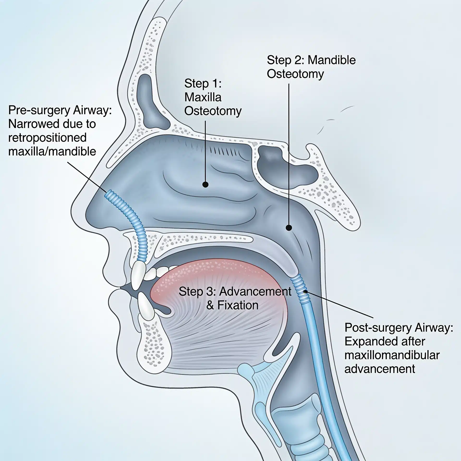
- Orthognathic Surgery:
- Used to realign the jaw or correct severe jaw deformities.
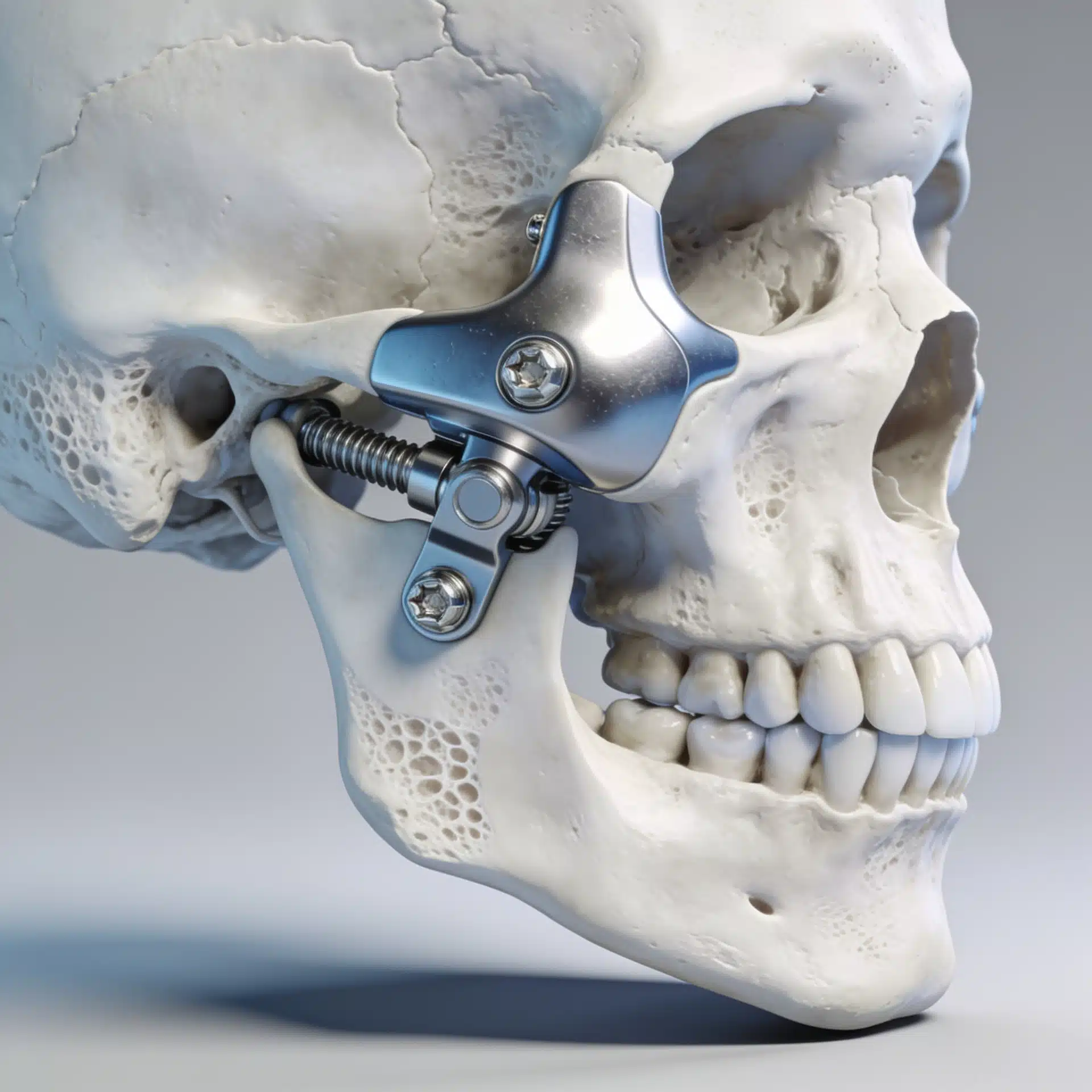
- Reconstructive Surgery:
- Rebuilding lost or damaged facial structures through bone grafts or artificial implants.
- Distraction Osteogenesis:
- Gradually lengthens bones using a mechanical device to correct deficiencies like micrognathia.
2. Non-Surgical Options
- Orthodontic Treatment:
Early intervention with braces or aligners can address dental alignment issues.
- Speech Therapy:
Particularly critical for conditions affecting oral and pharyngeal structures, such as cleft palate.
- Prosthetics:
Custom-made prosthetics, such as ears or noses, enhance function and appearance in select cases.
Early intervention with a clear treatment strategy improves both aesthetic and functional outcomes for patients.
Case Studies: Success Stories in Craniofacial Treatment
Case 1: Transforming Life with Early Cleft Lip Repair
A 1-year-old patient underwent a cleft lip repair surgery. Following the operation, the child experienced improved feeding mechanics and significantly fewer ear infections. Speech therapy further enhanced communication skills as the child aged.
Case 2: Cranial Vault Surgery for Craniosynostosis
A 6-month-old diagnosed with sagittal synostosis received cranial vault surgery to reshape their skull. By age two, cognitive development and skull growth were within the normal range, showcasing the importance of early surgical intervention.
These cases underscore the potential for highly successful outcomes when craniofacial conditions are managed promptly and effectively.
Future Directions in Craniofacial Treatments
Advancements in technology and research are expanding possibilities in craniofacial treatment. Promising developments include:
1. 3D Printing
From surgical planning to prosthetic creation, 3D printing is revolutionizing craniofacial surgery. Custom implants and anatomical models are improving precision and patient outcomes.
2. Stem Cell Therapy
Research into stem cells is revealing potential for regenerating bone and soft tissue, offering less invasive solutions in craniofacial reconstruction.
3. AI and Predictive Analytics
Artificial intelligence is playing an increasing role in diagnosing and predicting outcomes, allowing for better personalization of treatment plans.
4. Genetic Editing
CRISPR technology offers possibilities for addressing the underlying genetic causes of congenital craniofacial deformities.
These advancements hold immense potential for transforming the future of craniofacial care.
The Importance of Early Intervention
Managing craniofacial deformities requires a combination of medical expertise, advanced diagnostics, and innovative treatments. Early intervention is critical not only to mitigate physical complications but also to support emotional and psychological well-being.
Medical professionals have a unique opportunity to make a lasting impact on the lives of patients through timely and effective care. By staying informed about emerging trends and best practices, healthcare providers can ensure a brighter future for individuals living with craniofacial conditions.
Interested in exploring the latest tools for craniofacial diagnostics and treatment? Stay updated with our blog and resources to keep advancing your expertise.