Condylar Hyperplasia
Condylar hyperplasia (CH) is a generic term describing enlargement of the condyle. There are a number of different condylar pathologies that enlarge the mandibular condyle, with subsequent adverse effects on the morphology and function of the TMJ and mandible.
This may result in the development or worsening of a dentofacial deformity such as; mandibular prognathism (symmetric or asymmetric), and unilateral enlargement of the condyle, ramus, and body, facial asymmetry and malocclusion.
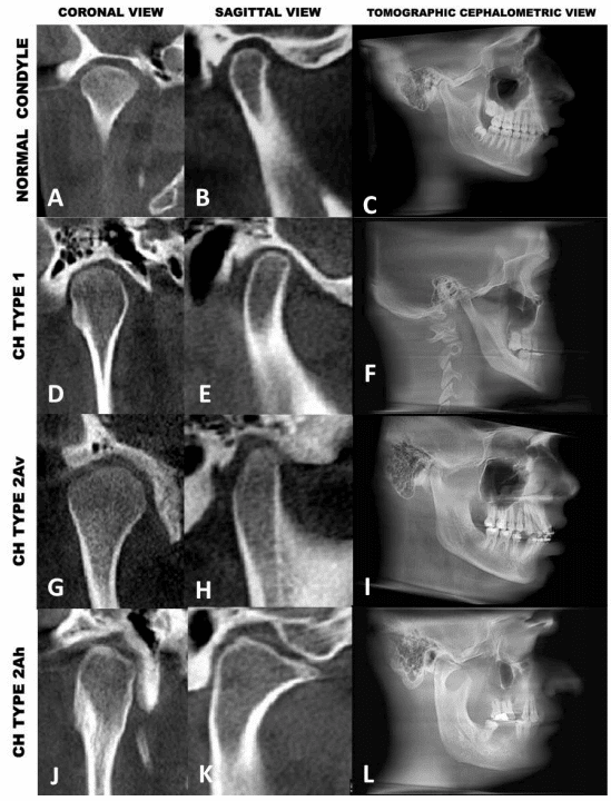
Dr. Wolford has developed a simple, but encompassing classification that will allow the clinician to better understand the nature of the various CH pathologies, progression, and treatment options that have proven to eliminate the pathological process and provide optimal functional and esthetic outcomes. The classification (Table 2 and Figure 29) also begins with the most common occurring form of Condylar Hyperplasia (CH) and progresses to the least common occurring form.
CH Type 1: This condition develops during puberty, is an accelerated and prolonged growth aberration of the normal condylar growth mechanism, is self-limiting but can grow into the 20’s, and can occur bilaterally (CH Type 1A) or unilaterally (CH Type 1B).
CH Type 2: These condylar pathologies can develop at any age (although 2/3s develop in the 2nd decade), are unilateral condylar vertical and/or horizontal over-growth deformities, and are the most common occurring mandibular condylar tumors; osteochondroma (CH Type 2A) and less common osteoma (CH Type 2B).
CH Type 3: These are other rare benign causing condylar enlargement.
CH Type 4: These are malignant conditions that can cause condylar enlargement.
The more common forms of CH (Types 1 and 2) will be presented relative to the clinical and radiographic findings, growth characteristics, effects on the jaws and facial structures, histology, and treatment considerations that are highly predictable in the elimination of the pathology and provide optimal treatment outcomes.
FIGURE 29 Description
A-C) normal TMJ with balanced joint spaces.
D-F) CH Type 1 with relatively normal condylar shape, elongated condylar head and neck, and narrow joint space related to thin articular disc or displaced disc. In the coronal view the condylar head is more rounded.
G-I) CH Type 2Av; an osteochondroma with a vertical growth vector without significant horizontal condylar enlargement or exophytic horizontal growth. This is a “young” osteochondroma with only about 3 years of growth.
J-L) CH Type 2Ah; an osteochondroma with horizontal (as well as vertical) enlargement of the condyle and exophytic outgrowth of the tumor. This tumor has been present for 6 years. Notice the significant increased vertical height of the mandibular body and ramus.
Mandibular Condylar Videos


