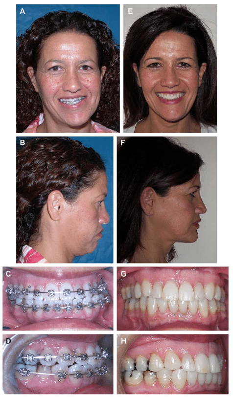
CH Type 2 – Condylar Hyperplasia Type 2; Osteochondroma Or Osteoma, Case 2
CH Type 2 is a unilateral over-development (enlargement) of the condyle usually caused by a benign tumor; osteochondroma (CH Type 2A) (CH Type 2B).
CH Type 2A and B will usually cause unilateral progressive vertical elongation of the mandible and compensatory down-growth of the maxilla, commonly develops a posterior open bite on the ipsilateral side, and a transverse cant in the occlusal plane, with ipsilateral side lower than the contralateral side.
CH Type 2 can occur at any age but 2/3rd of the time the onset is in the 2nd decade, it can usually be identified radiographically by an enlarged deformed condyle often with exophytic growth, increased width of the condylar neck, and increased vertical height of the mandible on the involved side. There are 2 basic radiographic and growth patterns:
- Vertical growth pattern (CH Type 2A) where the condyle elongates vertically without appreciable horizontal condylar enlargement (Figure 29: G-I) or exophytic growth extensions (Figure 29: J-L).
- Significant horizontal (and vertical) enlargement of the condyle (CH Type 2B) will have exophytic growth extensions of the tumor that can grow in any direction but more commonly in a forward projection.
MRI will show an ipsilateral enlarged, deformed condyle often with the disc in position but the contralateral side may have a displaced disc (75% of cases) and arthritic changes from the functional over-load to that joint as a consequence of the enlarged ipsilateral condyle.
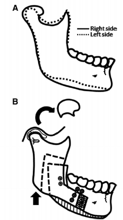
Our treatment protocol (Figure 33) for this TMJ pathology includes:
- Low condylectomy preserving the condylar neck;
- Recontour the condylar neck to function as a new condyle;
- Reposition the articular disc over the condylar stump, and contralateral disc if displaced with Mitek anchor technique;
- Orthognathic surgery usually including both jaws; and
- Vertical reduction of the inferior border of the mandible on the ipsilateral side if indicated to improve vertical facial balance with preservation of the inferior alveolar nerve.
Our studies 1 & 20 have shown that this technique is highly predictable to eliminate the TMJ pathology as well as maintain long-term skeletal and occlusal stability and facial balance.


