In the realm of medical advancements, MRI has long proven itself as an invaluable tool for providing detailed insights into the human body. But did you know that MRI technology is now revolutionizing TMJ replacement surgery? This groundbreaking approach is offering new hope for individuals suffering from temporomandibular joint (TMJ) disorders.
By harnessing the hidden power of MRI, surgeons are able to create custom-fit TMJ implants that closely mimic the patient’s natural joint structure. This innovative approach ensures a more precise fit, reducing the risk of complications and improving the overall success rate of the surgery.
In addition to enhancing surgical outcomes, MRI is also allowing surgeons to better evaluate and diagnose TMJ disorders before even stepping foot in the operating room. Through detailed imaging, doctors can accurately identify the specific issue, enabling them to develop more tailored treatment plans and ultimately provide patients with better long-term results.
With the help of advanced imaging techniques, the field of TMJ replacement surgery is being transformed, offering a brighter future for those suffering from this debilitating condition. Let’s explore how MRI is reshaping the landscape of TMJ treatment and the hope it brings to countless individuals around the world.
Challenges in traditional TMJ replacement surgery
Traditional TMJ replacement surgery has long been associated with challenges and limitations. The complex nature of the temporomandibular joint, combined with individual anatomical differences, makes achieving a precise fit for the implant a significant challenge. This often leads to a higher risk of complications, including implant failure, infection, and discomfort for the patient.
Additionally, traditional methods of diagnosing TMJ disorders have relied heavily on physical examinations and patient-reported symptoms, which may not provide a complete picture of the underlying issue. This lack of accurate diagnosis can result in suboptimal treatment plans and less successful outcomes for patients.
The role of MRI in TMJ replacement surgery
MRI has emerged as a game-changer in the field of TMJ replacement surgery. By utilizing advanced imaging technology, surgeons can now obtain highly detailed three-dimensional scans of the patient’s jaw, allowing for a more precise and personalized approach to treatment.
With the help of MRI, surgeons can evaluate the patient’s joint structure, identify any abnormalities or damage, and accurately measure the dimensions required for the custom-fit implant. This level of precision significantly reduces the risk of complications and improves the overall success rate of the surgery.
Furthermore, MRI provides invaluable information about the surrounding tissues, such as muscles, ligaments, and blood vessels. This allows surgeons to assess the overall health of the joint and plan the surgery accordingly, ensuring the best possible outcome for the patient.

Benefits of using MRI in TMJ replacement surgery
The use of MRI in TMJ replacement surgery offers a multitude of benefits for both patients and surgeons alike. Firstly, the ability to create custom-fit implants based on MRI scans ensures a more precise fit, reducing the risk of implant failure and the need for revision surgeries. This not only improves the patient’s quality of life but also reduces healthcare costs in the long run.
Additionally, MRI enables surgeons to visualize the joint in its entirety, providing a comprehensive understanding of the patient’s condition. This allows for more accurate diagnosis and the development of tailored treatment plans. By addressing the underlying cause of the TMJ disorder, rather than solely focusing on symptom management, patients can experience long-term relief and improved overall function.
Moreover, the non-invasive nature of MRI scans eliminates the need for exploratory surgeries, reducing patient discomfort and recovery time. This is particularly beneficial for individuals who may be hesitant to undergo invasive procedures or have underlying health conditions that make surgery more risky.
How MRI revolutionizes the diagnosis of TMJ disorders
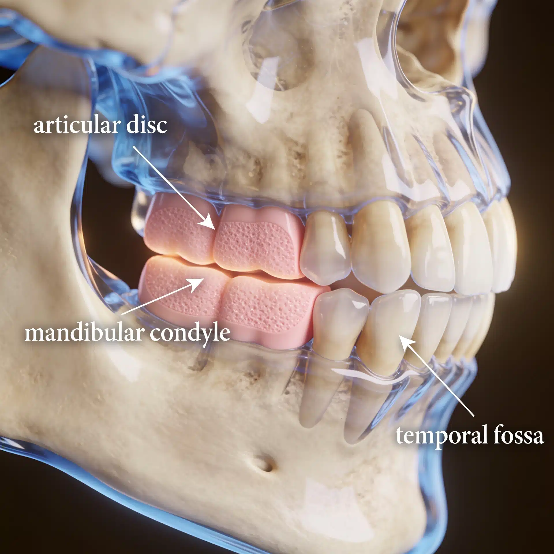
Diagnosing TMJ disorders accurately is crucial in determining the most appropriate treatment approach. MRI plays a pivotal role in revolutionizing the diagnosis of TMJ disorders by providing detailed imaging that captures the complexity of the joint and surrounding structures.
Through MRI scans, doctors can identify various factors contributing to TMJ disorders, such as joint degeneration, disc displacement, or abnormalities in the surrounding tissues. This allows for a more targeted treatment plan, addressing the specific issue rather than relying on generalized approaches.
Furthermore, MRI can detect early signs of TMJ disorders, enabling proactive intervention and preventing the condition from progressing to a more severe stage. Early diagnosis and intervention increase the likelihood of successful treatment outcomes and minimize the need for extensive surgical interventions.

Preparing for TMJ replacement surgery with MRI
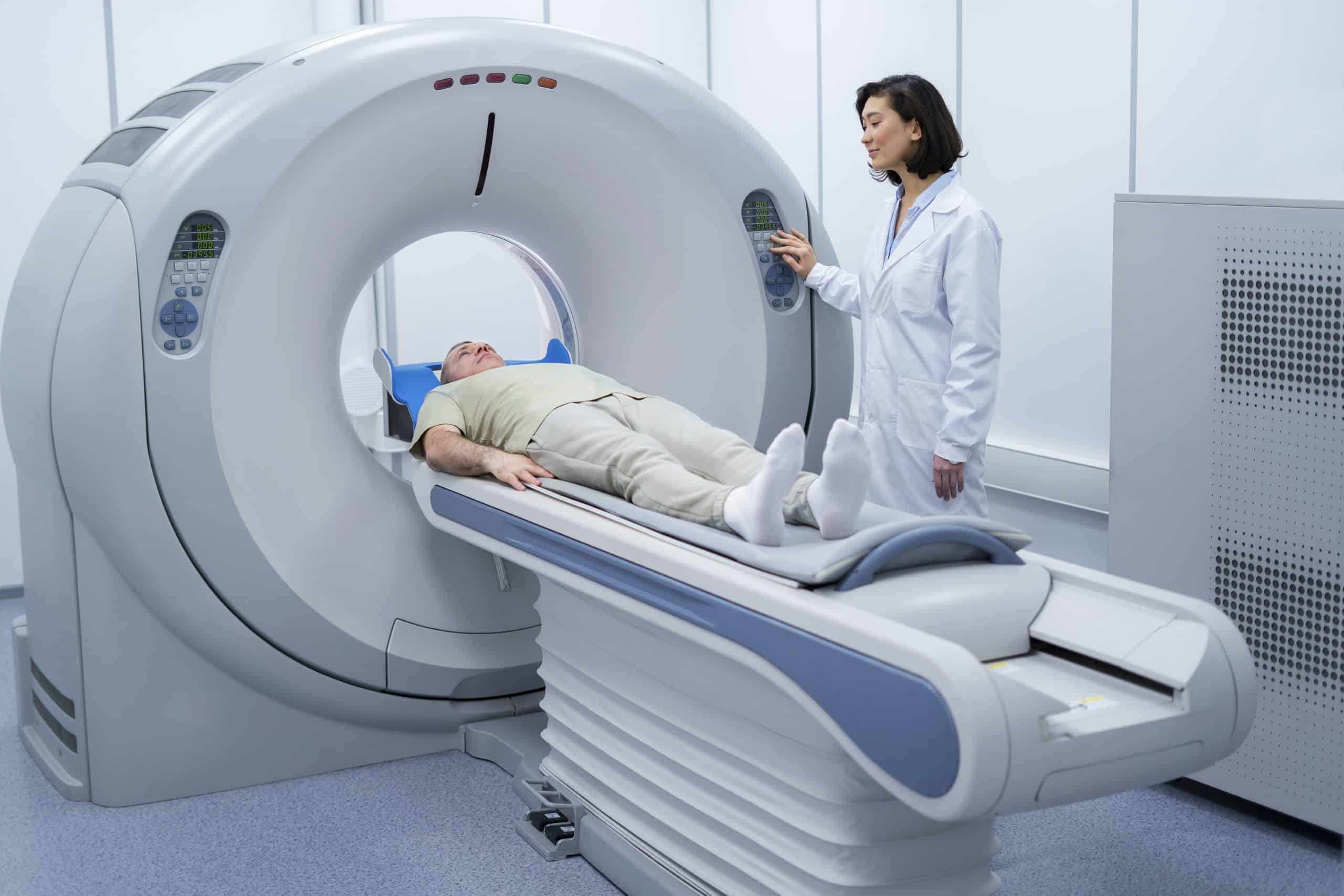
Before undergoing TMJ replacement surgery, patients will typically undergo a series of MRI scans to assess the joint’s condition and plan the surgical procedure. The preparation process involves several steps to ensure the best possible outcome.
Firstly, the patient will be scheduled for an MRI appointment, during which they will be positioned in a specialized scanner that captures detailed images of the jaw and surrounding structures. The scanning process is painless and non-invasive, requiring the patient to remain still for a specific duration.
Once the MRI scans are complete, the images will be processed and analyzed by a team of specialists, including radiologists and surgeons. These experts will carefully evaluate the scans to assess the joint’s health, identify any abnormalities, and plan the surgical approach accordingly.
The procedure of TMJ replacement surgery with MRI guidance
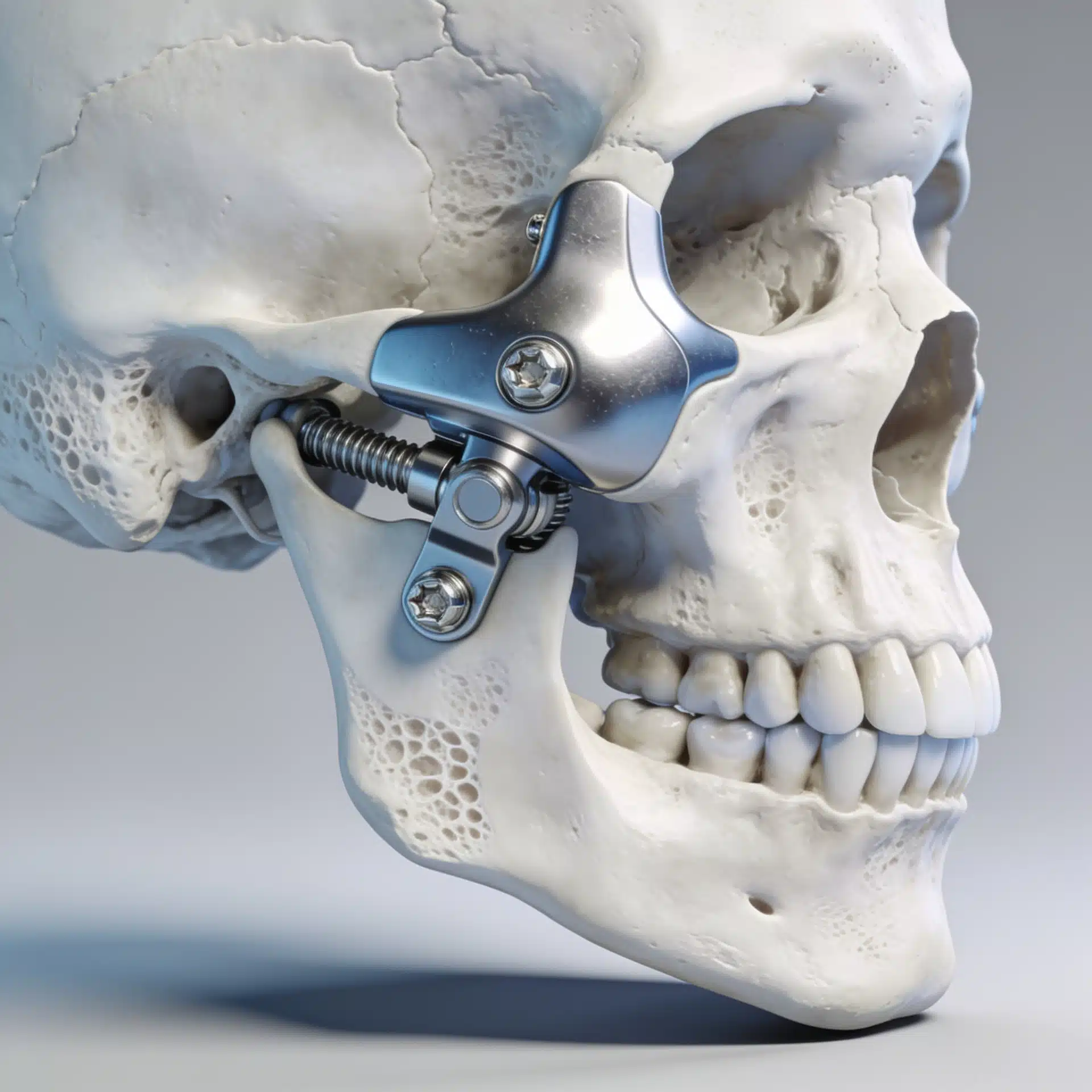
TMJ replacement surgery with MRI guidance involves a comprehensive and precise surgical procedure to replace the damaged joint with a custom-fit implant. The surgery itself is typically performed under general anesthesia and involves several key steps.
Firstly, the surgeon will make an incision in the skin, providing access to the TMJ. The damaged joint components, including the disc and any degenerated or damaged bone, will be carefully removed. This creates a clean slate for the implantation process.
Next, the surgeon will utilize the MRI scans to guide the placement of the custom-fit implant. The three-dimensional imaging allows for a precise fit, ensuring optimal alignment and reducing the risk of post-operative complications.
Once the implant is securely positioned, the surgeon will close the incision, and the patient will be moved to a recovery area. Post-operative care and rehabilitation will be necessary to facilitate healing and restore normal function to the jaw.

Recovery and post-operative care for TMJ replacement surgery
Recovering from TMJ replacement surgery is a gradual process that requires patience and compliance with post-operative care instructions. The initial days following the surgery are crucial for proper healing and avoiding complications.
Patients will likely experience some swelling, discomfort, and limited jaw movement immediately after the surgery. Pain medication and ice packs can help alleviate these symptoms. Soft foods and a liquid diet may be recommended during the initial recovery period to avoid placing excessive strain on the jaw joint.
Physical therapy and jaw exercises are often prescribed to improve jaw mobility and strengthen the surrounding muscles. Following these exercises diligently is essential for restoring normal function and preventing stiffness or long-term complications.
Regular follow-up appointments with the surgeon will be scheduled to monitor the progress of healing and assess the success of the surgery. These appointments provide an opportunity for any concerns or issues to be addressed promptly, ensuring the best possible outcome for the patient.
Success stories of patients who underwent TMJ replacement surgery with MRI
The success of TMJ replacement surgery with MRI guidance is evident in the countless stories of individuals who have regained their quality of life after suffering from TMJ disorders. These success stories highlight the transformative power of advanced imaging techniques in the field of TMJ treatment.
Patients who underwent TMJ replacement surgery with MRI have reported a significant reduction in pain, improved jaw function, and an overall improvement in their ability to carry out daily activities. Many individuals have been able to regain their ability to eat, speak, and laugh without discomfort, allowing them to enjoy a better quality of life.
Furthermore, the precision and accuracy of MRI-guided TMJ replacement surgery have led to a reduced need for revision surgeries or additional interventions. This not only minimizes the physical and emotional burden on the patient but also reduces healthcare costs and resources.
Conclusion: The future of TMJ replacement surgery with MRI
The integration of MRI technology into the field of TMJ replacement surgery has ushered in a new era of precision and personalized treatment. By harnessing the hidden power of MRI, surgeons can now offer patients custom-fit implants that closely mimic their natural joint structure, enhancing surgical outcomes and improving the overall success rate of the procedure.
Moreover, MRI enables accurate and comprehensive diagnosis of TMJ disorders, allowing for tailored treatment plans that address the underlying cause of the condition. This proactive approach ensures better long-term results and minimizes the need for extensive surgical interventions.
As technology continues to advance, the future of TMJ replacement surgery with MRI holds even more promise. Further refinements in imaging techniques and surgical approaches will continue to enhance patient outcomes and bring new hope to those suffering from TMJ disorders.
With each success story and research breakthrough, the power of MRI in revolutionizing TMJ replacement surgery becomes increasingly evident. Through the collaboration of medical professionals, researchers, and technological advancements, the field of TMJ treatment is evolving, offering a brighter future for individuals worldwide.